Embarking on a journey into anatomy at home is now achievable, offering hands-on learning that significantly boosts comprehension and retention. This guide will help you build your own models, turning learning into a fun and engaging process.
The Value of Hands-On Learning
Hands-on learning in anatomy dramatically enhances understanding, moving beyond rote memorization to a deep, practical grasp of complex structures. Engaging with DIY models, like creating a shaking incubator or a smartphone microscope, facilitates a more intuitive comprehension of anatomical concepts. This method promotes active learning, enabling students to visualize and interact with the subject matter, leading to better retention and recall. Such learning also fosters problem-solving skills, as students navigate the challenges of model construction, reinforcing their understanding of anatomy through practical application. Moreover, it encourages creativity and innovation, allowing for personalized learning experiences that cater to individual learning styles and preferences, making anatomy more accessible and enjoyable. This active engagement also increases motivation, making study sessions much more productive.

DIY Anatomy Models
Creating your own anatomy models at home is a fantastic way to explore and understand complex structures. These hands-on projects make learning more engaging and effective.
Building a Simple Shaking Incubator
Transforming an old record player into a functional shaking incubator is a creative way to explore biological processes at home. This project involves repurposing a vintage device into a valuable piece of lab equipment. By carefully securing a platform to the turntable, you create a stable base for culture flasks or Petri dishes. This shaking action is critical for even distribution of nutrients and oxygen, fostering optimal growth. Ensure the speed is adjustable to suit various experiments. Remember safety first; always supervise your experiments and properly clean your DIY incubator. This method provides a hands-on understanding of laboratory techniques without needing expensive equipment. It’s a perfect example of how everyday items can be used in a scientific setting, making science more accessible.
Creating a Smartphone Microscope
Transform your smartphone into a powerful microscope using readily available materials, allowing you to explore the microscopic world with ease. By attaching a simple lens, often sourced from a laser pointer or inexpensive toy, to your phone’s camera, you can create a functional microscope. Securing the lens with tape or a small clamp ensures stability. Proper lighting is vital for clear images, so consider using an LED flashlight or a bright lamp. This DIY microscope is perfect for observing cell structures and other tiny details. The convenience of using your smartphone lets you capture and share images instantly, making it an excellent tool for learning. It’s an innovative and affordable method to explore the world of anatomy at a microscopic level, encouraging curiosity and discovery.
Preparing for Anatomy Lab Practicals
Effective preparation is key for success in anatomy lab practicals. This section explores strategies for studying and utilizing visual aids, which are essential for mastering anatomical concepts and procedures.
Effective Study Strategies
Mastering anatomy requires a blend of memorization and recall skills. Engaging with new vocabulary through various methods, like grouping key terms and matching structures, is crucial. Fresh memory is vital, so review topics just before practicals, such as the flexors of the forearm. Utilize resources like YouTube channels for organ preparation and real bones for studying skeletal structures. Discussing with peers is also helpful. Remember, time management is essential, especially when juggling multiple courses. Start with a readiness quiz to ensure preparation. Divide into groups during labs to answer questions collectively. Allocate time specifically for anatomy, potentially reducing other commitments. Concentrate on the most challenging topics for the best results.
Utilizing Visual Aids
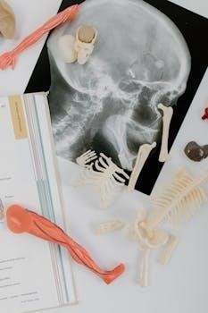
Visual aids are paramount in anatomy study, helping to understand complex structures. Explore resources like 3D models and anatomical charts to visualize the body’s systems. Use online tools and videos, such as those offered by 3D4Medical, to gain a better perspective. Consider using anatomical models as hands-on learning tools. Creating your own gelatin models can be a fun and effective way to grasp spatial relationships. X-ray images can provide a clear understanding of bone structure, including fractures and epiphyseal plates. Moreover, diagrams and illustrations can simplify intricate anatomical concepts. Remember to actively engage with visual resources, not just passively observe them. Make sure you review the relationships that are difficult to understand using visual aids. This active method enhances comprehension and retention.

Essential Equipment for a Home Lab
Setting up a home anatomy lab requires careful planning and sourcing of materials. A safe workspace and proper tools are necessary for successful experiments, ensuring a productive study experience.
Sourcing Necessary Materials
Gathering the right materials is a key step in setting up a home anatomy lab. Begin by identifying the specific needs of your planned experiments and models. Consider what is required for the type of study you want to accomplish, whether it’s building a model of the hand or simply examining preserved specimens. You may find that many items can be obtained from local craft stores, online retailers, or even repurposed from household items. For instance, modeling clay, simple tools, and even gelatin can prove very useful. Don’t forget to source safety goggles and gloves for protection, and look for preserved specimens, if needed, from educational supply companies. Always ensure materials are safe, non-toxic, and suited for hands-on learning. A well-planned material list is essential for a successful lab experience.
Setting Up a Safe Workspace
Creating a safe workspace is paramount for any home anatomy lab. Begin by selecting a designated area, ideally near a sink to aid in cleanup. Ensure the area is well-lit and has proper ventilation. Cover the work surface with protective material to prevent damage and ensure easy cleaning. Keep all materials organized and easily accessible to prevent accidents. Have a first-aid kit readily available for minor injuries. Always use personal protective equipment, such as gloves and eye protection, when handling specimens or chemicals. Establish clear safety guidelines with everyone using the lab, especially if children are involved. Properly dispose of all waste materials. A well-organized and safe space will help you stay focused and prevent unfortunate accidents, letting you enjoy your lab experience.

Working with Specimens
When working with specimens, careful dissection techniques are essential for learning. Remember to follow all ethical guidelines, and always treat specimens with the utmost respect while studying.
Dissection Techniques
Mastering dissection techniques is crucial for a successful home anatomy lab. Start by ensuring you have proper tools, such as a scalpel, scissors, and forceps, all in good condition. Begin with a shallow incision, carefully separating layers of tissue, taking note of the different textures and structures you encounter. It is essential to proceed methodically, always referencing your anatomy guide to accurately identify each part. Use gentle movements to avoid tearing or damaging delicate structures. Remember to separate nerves from connective tissue without harming them, and be precise in your cuts. Proper technique will allow you to understand the spatial relationships between various organs and tissues. Do not hesitate to ask an experienced person to show you their methods for challenging tissue separations, if you can. The goal of dissection is to reveal, not destroy, the specimen’s anatomical architecture.
Ethical Considerations
When engaging in home anatomy labs, ethical considerations must be paramount. If using animal specimens, ensure they were sourced responsibly and humanely, ideally from reputable suppliers who follow ethical guidelines. Treat all specimens with respect, understanding they were once living beings. Avoid unnecessary damage or waste during dissection. Consider proper disposal methods for specimens and related materials, complying with local regulations. It is important to acknowledge the educational value the specimen offers and approach your work with a sense of responsibility. If the home lab involves human models or simulations, it’s crucial to maintain privacy and respect. Be mindful of the sensitivity involved in working with anatomical subjects and always prioritize ethical practices throughout your home lab experience. Remember that the goal is educational enrichment, not entertainment or disregard for life.

Advanced Home Lab Projects
Elevate your learning with advanced projects, such as building a robotic hand model or creating gelatin anatomy models. These projects offer deeper understanding through hands-on creation and exploration of complex structures.
Building a Robotic Hand Model
Creating a robotic hand model is an engaging project that allows for an in-depth exploration of the complex anatomy of the human hand. This project goes beyond basic memorization, offering a practical understanding of how tendons, ligaments, and muscles work together to facilitate movement; By constructing each component, you will gain a deeper appreciation for the intricate design of the hand. Start with a basic design using readily available materials such as cardboard, string, and simple actuators. The process involves carefully replicating the movements of fingers and thumbs, providing an interactive learning experience. This project enhances your understanding of biomechanics and offers a hands-on approach to learning complex anatomical principles. It is a challenge that combines engineering and anatomy, perfect for those looking to take their studies to the next level. Remember to document each step to fully grasp the process.
Creating Gelatin Anatomy Models
Constructing anatomy models from gelatin offers a unique, hands-on method to visualize internal structures. This approach is not only cost-effective but also allows for a tactile learning experience, making it ideal for exploring the spatial relationships between organs and tissues. Start by preparing a clear gelatin base; then, you can embed various items to represent anatomical structures, such as colored candies, strings, or small plastic components. The key is to ensure that all embedded materials are clearly visible and allow for easy identification. This method transforms abstract concepts into a concrete form, enhancing your spatial understanding. This is also a fantastic way to demonstrate organ systems and how they interact within the body, providing a three-dimensional view. This project also offers a great way to make learning fun and engaging.

